
What is Gangliosidosis GM1 Type I?
GM1-gangliosidosis type I (generalized familial gangliosidosis, neurovisceral lipidosis, b-galactosidase deficiency) is characterized by an autosomal recessively inherited deficiency of the enzyme GM1-b-galactosadase. The activity of this enzyme in the brain, liver and fibroblasts of the patient’s skin is reduced to 0.1% of normal. More than 100 cases of the disease have been described.
Causes of Gangliosidosis GM1 Type I
The basis of the disease is a mutation of the structural gene encoding the monomeric polypeptide A1GM1-b-galactosidase and localized on the short arm of the 3rd chromosome (p12q21). This defect impairs the cleavage of terminal galactose from GM1 ganglioside, which is deposited in large quantities in the gray matter of the brain and liver.
Symptoms of Gangliosidosis GM1 Type I
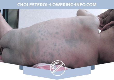
GM1-gangliosidosis is detected at birth or soon after it. Poor appetite, weakness of sucking and screaming, insufficient weight gain, swelling of the lower extremities, muscular hypotonia and low activity of the child (calm, sleep most of the time). Hepatosplenomegaly is observed from the first months of life. Often there are dorsolumbalny scoliosis, unsharp increase in the size of the joints, shortening and thickening of the fingers (brachydactyly). Radiographically determined multiple dysostosis. Characterized by frequent bronchopneumonia. Clonic-tonic convulsions develop. Approximately 50% of patients with early-developed GM1 gangliosidosis have cherry-red spots on the fundus of the yellow spot, resulting from the deposition of gangliosides in retinal cells. If the child has lived to 6 months, then he has a characteristic appearance (protruding frontal bumps, sunken bridge, large, low-lying ears, gum hypertrophy, macroglossia, swelling of the face).
By 8-9 months, the child is not sitting, not crawling, his movements are uncoordinated, muscular hypotonia is replaced by hypertonus, tendon reflexes increase. By the end of the first year of life, deafness, blindness, spastic tetraplegia, a lack of response to the environment, decerebration rigidity develops at the terminal stage.
Children with GM1 gangliosidosis usually die at the age of 2–3 years from recurrent bronchopneumonia.
Diagnosis of Gangliosidosis GM1 Type I
An autopsy reveals the expansion of the ventricles of the brain and its atrophy as a result of the death of neurons. Histologically detected frothy histiocytes in the bone marrow, liver, spleen, lymph nodes, etc.
The diagnosis of GM1-gangliosidosis is confirmed when determining the activity of b-galactosidase in leukocytes and cultured fibroblasts. GM1-gangliosidosis type I should be differentiated from mucopolysaccharidosis type I – Hurler syndrome, Niemann-Pick disease and I-cell disease. GM1-gangliosidosis type I occurs at an earlier age than Gurler syndrome and Niemann-Pick disease. The latter is characterized by clouding of the cornea, damage to the bones, a less pronounced change in facial features.
Treatment of Gangliosidosis GM1 Type I
Specific therapy is absent. Methods of substitution therapy with b-galactosidase (purified or encapsulated in liposomes) are being developed.